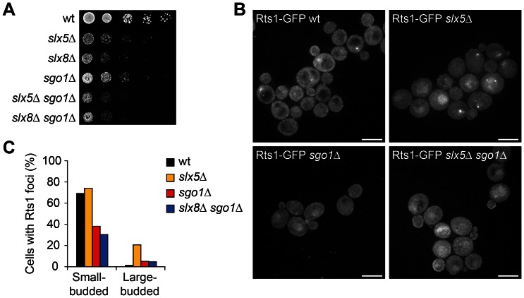
Figure 5. Rts1 foci are partially Shugoshin (Sgo1)-dependent.
(A) Growth rate assay of cells spotted in five-fold serial dilutions on YPD plates. Images are after two days growth at 25°C. (B) Live cell fluorescence microscopy of asynchronous wt, slx5Δ, sgo1Δ and slx5Δ sgo1Δ cells expressing Rts1-GFP. Scale bars, 5 µm. (C) Quantification of Rts1 foci in cells, shown in (B). Quantification is based on an asynchronous cell population (n = 89–203), that was morphologically divided in a small-budded and large-budded cell population.

