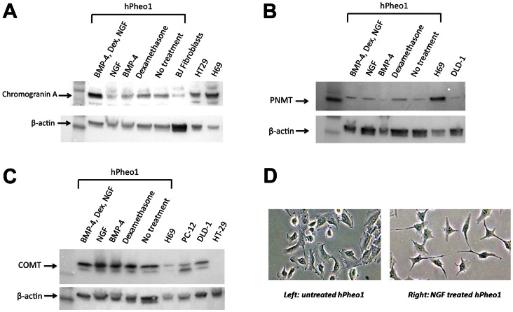
Figure 2. Differentiation of hPheo1 cells.
A. Western blot for chromogranin A shows expression more prominent after differentiation treatment. B. Western blot for PNMT shows expression more prominent after differentiation treatment. C. Presence of COMT in hPheo1 cells is seen, whether treated with differentiating factors or not. D. Neurite formation in hPheo1 cells occurs after treatment with Nerve Growth Factor (NGF).

