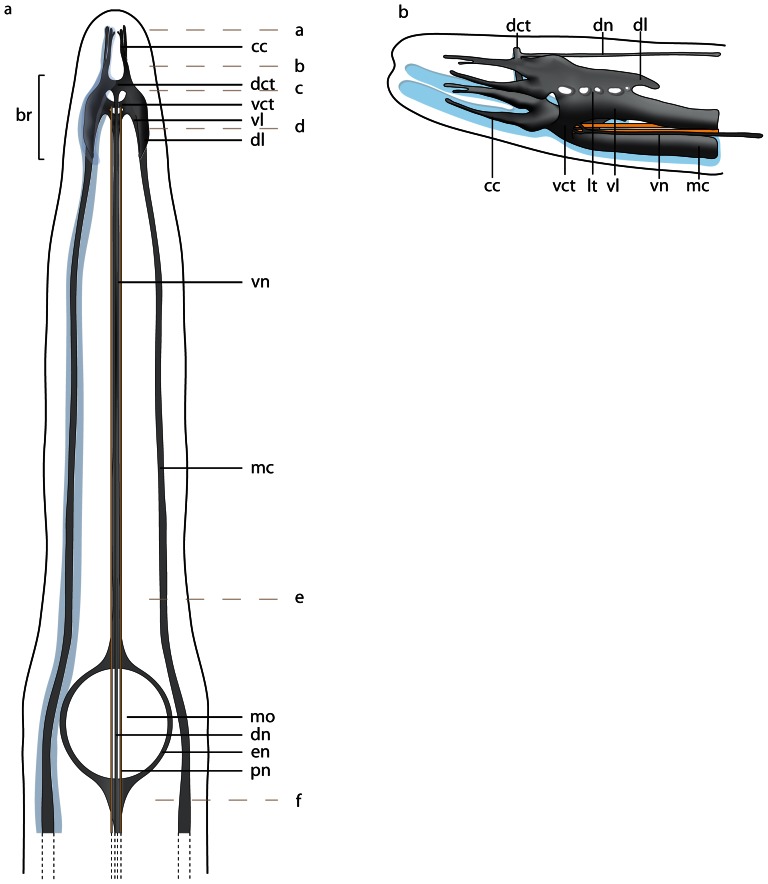
Figure 4. Procephalothrix filiformis, schematic drawings of the central nervous system based on 3D-reconstruction of 501 aligned 0.5 µm sections, dorsal (a) and lateral (b) view.
a: The nervous system is composed of neuropil (np, gray) which may be surrounded by cell somata (cs, blue). Cephalic cords (cc) are circular arranged around the head of the animals. The paired proboscidial nerves (pn, yellow) originate from the ventral commissural tract (vct). A dorsal nerve (dn) originates from the dorsal commissural tract (dct), a ventral nerve (vn) from the ventral commissural tract. The branching esophageal nerves (en) originate from the ventral nerve and surround the mouth opening (mo). The lateral medullary cords (mc) originate in the ventral lobes of the brain (br). Letters on the right (a–f) refer to the histological sections in figure 5 . The nerve plexus originating in the dorsal nerve (see fig. 5e ) is omitted. b: Central nervous system lateral view. The dorsal lobe (dl) of the brain and the ventral lobe (vl) of the brain are connected by several lateral tracts (lt).

