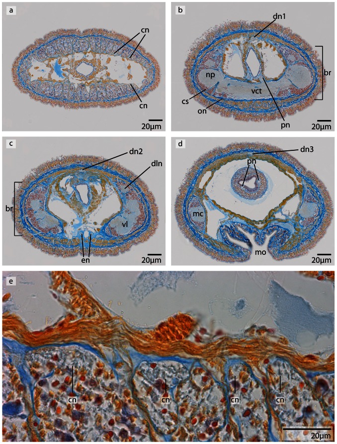
Figure 12. Carinoma mutabilis, light micrographs of Azan stained transverse sections of the brain.
a: The cephalic nerves (cn) are circularly arranged around the inner margins of the head. b: The brain (br) is composed of a central neuropil (np) surrounded by cell somata (cs). The whole brain is encircled by an outer neurilemma (on). The two proboscidial nerves (pn) originate from the ventral commissural tract (vct). c: A dorsal nerve (dn) arises from the dorsal commissural tract, the esophageal nerves (en) originate from the ventral commissural tract, the brain is in its posterior part divided into a dorsal (dl) and ventral (vl) lobe. Dorso-lateral nerves (dln) arise from the dorsal commissural tract and merge with the dorsal lobes. d: The ventral lobes are confluent with the lateral medullary cords (mc), two proboscidial nerves are present (pn). Note the different positions of the dorsal nerve (dn1–dn3). e: Higher magnification of the anterior head region, showing the location of the cephalic nerves (cn). dn dorsal nerve, mo mouth opening.

