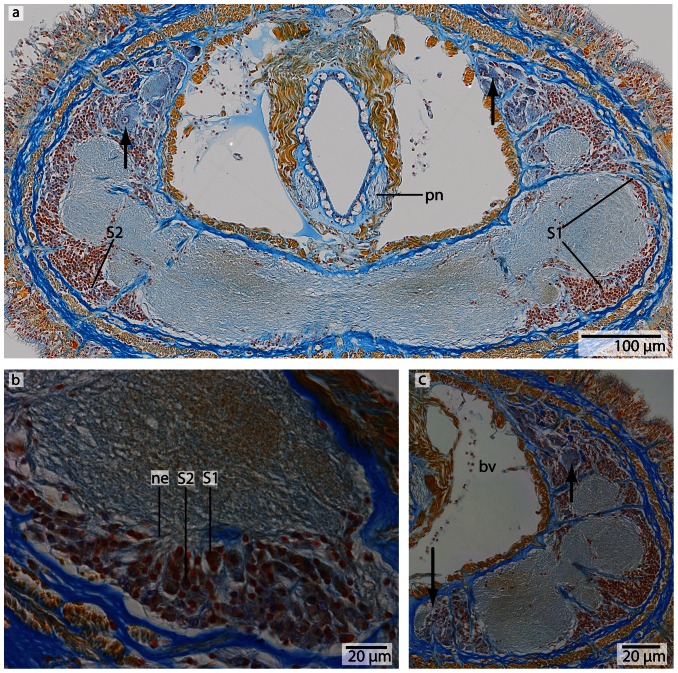
Figure 13. Carinoma mutabilis, light micrographs of Azan stained neurons.
a: Transverse section, overview of the brain, position of the two different types of neurons (S1–S2). Another type of cells is located in the dorsal tip of the brain (arrow). b: Higher magnification of somata of type 2 neurons (S2). The nuclei of S2 dye purple. The neurites (ne) of the brain neuropil branch into the somata layer. c: In the dorsal and ventral tip of the brain, cells with red-stained nuclei and very prominent cell bodies (arrows) are found. Note the close association of these cells with the blood vessels (bv). These cells resemble glandular cells. pn proboscis nerve.

