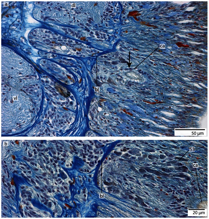
Figure 20. Tubulanus superbus, light micrographs of Azan stained transverse sections of the brain and cerebral organ.
a: The sensory cells (arrow) surrounding the canal (ca) of the cerebral organ are connected to the dorsal lobe (dl) of the brain. The canal ends anterior to the basal lamina (bl) of the epidermis. b: The cells adjacent to the cerebral organ are type 1 brain cells. vl: ventral lobe.

