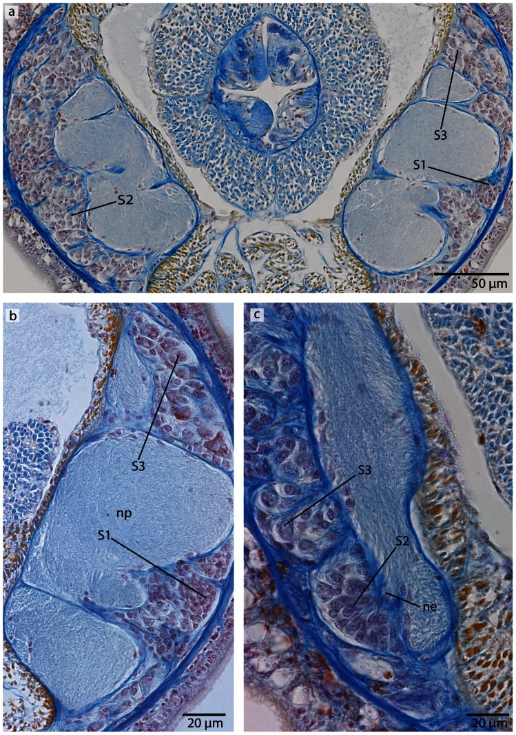
Figure 23. Callinera grandis, light micrographs of Azan stained neurons.
a: Transverse section, overview of the brain; position of the three different types of neurons (S1–S3); all nuclei stain purple. b: Higher magnification of somata of type 1 and 3 neurons (S1, S3). Cell bodies are circular; those of S1 are arranged in clusters, those of S3 are most prominent. c: Higher magnifcation of somata of type 2 and 3 neurons. S2 cell bodies are pear shaped and slightly enlarged. The neurites (ne) of the brain branch into the cell somata layer. np neuropil.

