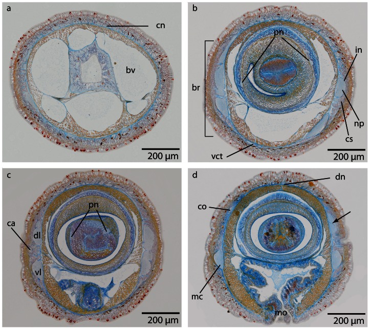
Figure 25. Carinina ochracea, light micrographs of Azan stained transverse sections of the brain.
a: Frontal region showing the circularly arranged cephalic nerves (cn). bv: blood vessel. b: The brain (br) is composed of a central neuropil (np) which is surrounded by cell somata (cs). A ventral commissural tract (vct) connects the two halves of the brain, and an outer neurilemma (on) encloses the whole brain. (pn): proboscidial nerves. c: The canals (ca) of the cerebral organs open laterally, where the brain divides into a dorsal (dl) and ventral (vl) sections. Two proboscidial nerves (pn) are present. d: The canals of the cerebral organs (co) end dorsally. A dorsal nerve strand (dn) arises from the dorsal commissural tract, the ventral part of the brain is confluent with the lateral medullary cords (mc). Note the ecm (arrow) branching into the neuropil of the brain. mo mouth opening.

