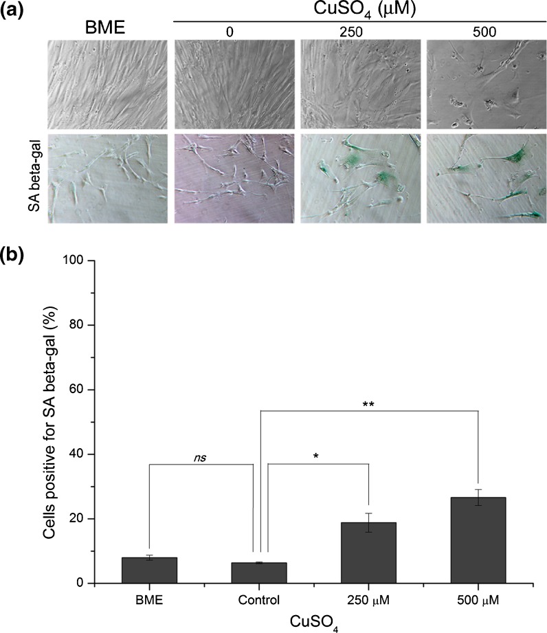
Fig. 2.
Cell morphology and senescence-associated β-galactosidase activity detection on fibroblasts exposed to 250 or 500 μM of copper sulfate. a Wi-38 HDFs exposed to 250 or 500 μM CuSO4 presented enlarged cellular volume and altered shape, resembling the typical senescent phenotype that normally appears on replicatively senescent fibroblasts. b SA β-gal-positive cells increased to 19% and 27% in cells exposed to 250 and 500 μM copper sulfate, respectively, when compared with control cells (6%). Data are expressed as mean ± SEM from three independent experiments. *p < 0.05; **p < 0.01 and ns non-significant, when compared with control

