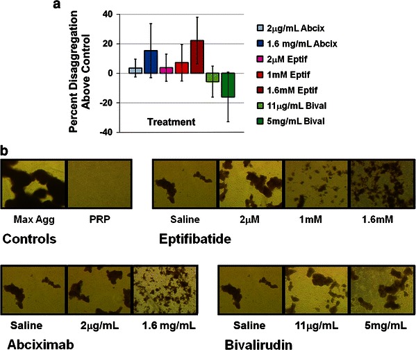
Fig. 2.

Disaggregation of aged platelet aggregates following exposure to GPIIb–IIIa antagonists (abciximab or eptifibatide) or bivalirudin. a Percent disaggregation (normalized to vehicle control) at 15 min post-drug addition. Results are expressed as mean ± SD (error bars), n = 4. b Bright field microscopy images of platelet aggregates fixed with paraformaldehyde at 15 min post-drug addition (40× magnification). Images are from a single donor that was representative of an n = 4
