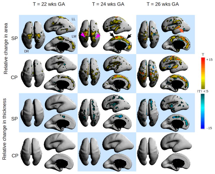
Fig. 2.
Spatiotemporal patterns of variational growth between the cortical plate (CP) and subplate (SP) during early fetal development. Warm colors indicate significant, relative increase in area or thickness component of growth with time and cool colors indicate statistically significant reductions in area or thickness component. DR = Dorsal view, LL = left lateral view and LM = left medial view. Changes are bilateral unless noted otherwise.

