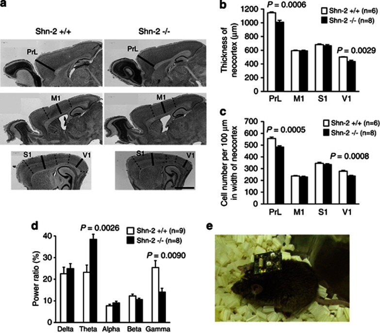
Figure 5.
Abnormalities in the cortex of Shn-2 KO mice. (a, b) The cortex of Shn-2 KO mice was thinner than that of wild-type mice. Cortical cell density was also reduced in the prelimbic cortex (PrL) and primary visual cortex (V1) in Shn-2 KO mice (c). (d) Theta band power increased and gamma power decreased in Shn-2 KO mice. (e) A mouse with the Neurologger, a head-mounted EEG data logger device. M1, primary motor cortex; S1, primary somatosensory cortex. Scale bar indicates 1 mm (a).

