Figure 4.
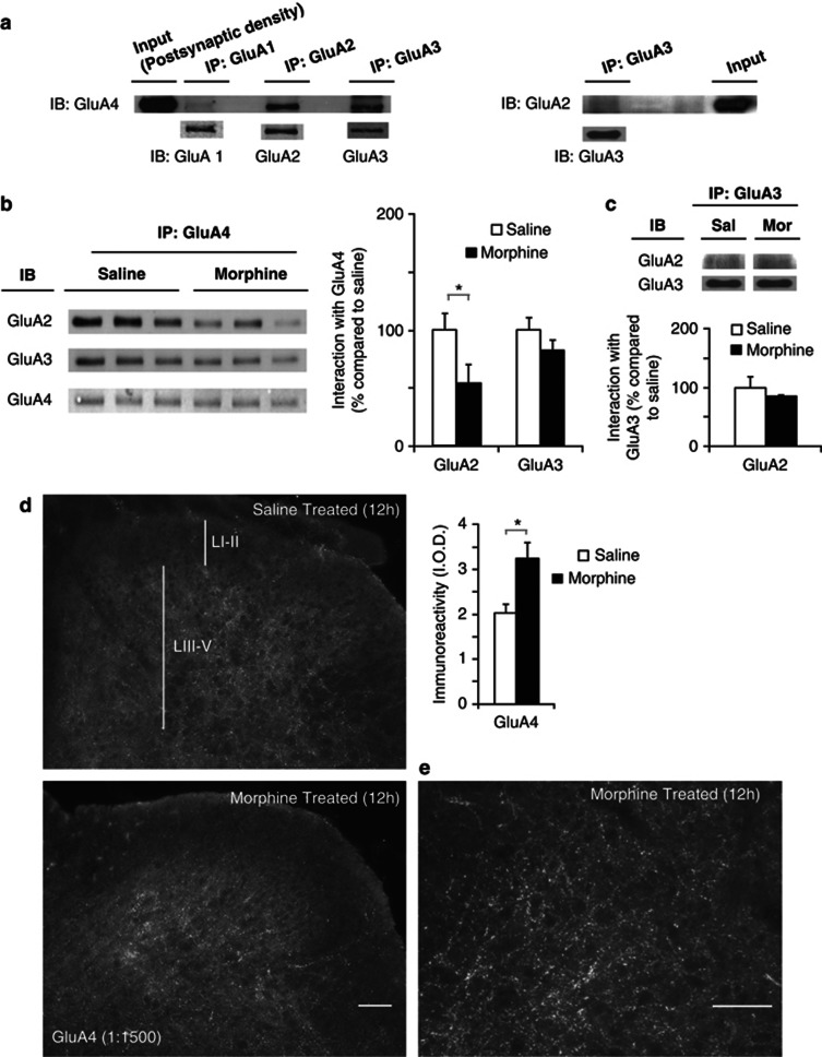
Composition of postsynaptic GluA4-containing AMPAR is altered after morphine treatment. The increase in GluA4-containing AMPAR is located in laminae III–V of spinal cord dorsal horn. (a) Basal composition of GluA4-containing AMPARs in the postsynaptic density. Co-immunoprecipitation of GluA1, GluA2, and GluA3 with GluA4 shows that in naive animals, GluA4 is associated with GluA2 and GluA3, however, association between GluA4 and GluA1 is not detected. GluA2 exhibits weak association with GluA3. (b) The proportion of GluA4 associated with GluA2 is decreased 12 h after morphine (▪), but not saline (□) treatment, whereas the association between GluA4 and GluA3 remains unaltered. Quantification was performed relative to GluA4 levels detected in the pull down. Values are expressed as mean %±SEM compared with the saline group, n=3 samples per group, *p⩽0.05, Mann–Whitney U-test. (c) The proportion of GluA2 associated with GluA3 did not change after morphine treatment. Quantification was performed relative to GluA3 levels detected in the pull down. (d) The increase in GluA4 expression is located in dorsal horn laminae III–V, morphine (▪) saline (□), values represent IOD (means±SEM) n=12, *p<0.01, unpaired t-test. (e) Representative higher magnification image of GluA4 staining in dorsal horn laminae III–V from morphine-treated mouse. Immunostained puncta outline cell bodies in these laminae, consistent with the receptor having a postsynaptic location. Scale bars=10 μm.

