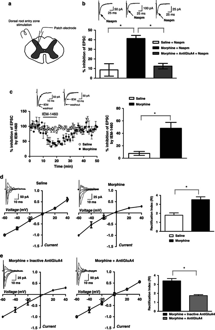
Figure 5.
Whole-cell patch-clamp recordings indicate morphine-induced insertion of GluA4-containing AMPAR in laminae III–V of spinal cord dorsal horn. (a) The spinal cord slice cartoon shows positioning of the electrodes. (b) AMPAR-mediated EPSCs show increased sensitivity to naspm in spinal cord slices from morphine-treated animals, which is reversed by infusion of GluA4 antibody. Sample traces (average of 20 trials) were recorded before and after naspm application (100 μM). Histograms show an average percentage inhibition (means±SEM) of EPSCs. (c) The effect of another Ca2+-permeable AMPAR blocker, IEM-1460, in slices from morphine-treated mice is similar to naspm. The amplitude of AMPAR-mediated EPSCs is plotted against time (left panel) and the histogram shows average percentage inhibition (right panel). Sample traces (average of 20 trials) were recorded before and after IEM-1460 application (50 μM). (d) Current–voltage relationships obtained by plotting AMPAR-EPSC amplitude as a function of holding potential (average I–V curves are shown). In saline-treated animal, I–V curves are almost linear showing very little contribution of Ca2+-permeable AMPAR. I–V curves obtained from a morphine-treated animal shows inward rectification – an indicator of Ca2+-permeable AMPAR insertion. Average RI calculated as EPSC (−60/+40 mV) is increased by morphine compared with saline. (e) I–V curve in morphine-treated animal is not affected by heat-inactivated anti-GluA4 included in the patch solution. Anti-GluA4 included in patch solution restores linearity of I–V curve from a morphine-treated animal. Average RI recorded from the slices of morphine-treated mice in the presence of heat-inactivated anti-GluA4 is similar to RI recorded without antibody. Anti-GluA4 included in the pipette completely reverses RI to the levels recorded in saline-treated mice, showing that Ca2+-permeable AMPARs inserted at the synapse after morphine consist of GluA4 subunits. *p<0.05.

