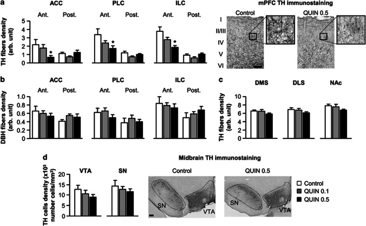
Figure 1.
(a) Density of tyrosine hydroxylase (TH) immunostained fibers in the anterior (Ant.) and posterior part (Post.) of the mPFC (including ACC, PLC, and ILC) in vehicle- (control, white bars; n=9) or quinpirole-treated rats (QUIN 0.1, gray bars; n=12/QUIN 0.5, black bars; n=10). Right microphotographs show a representative TH immunostaining in superficial and deep layers (I–VI) of the PLC for control and QUIN 0.5 rats (scale bar=200 μm). Insets represent higher magnification of delineated areas. (b) Density of dopamine-β-hydroxylase (DBH) immunostained fibers in the anterior and posterior part of the mPFC. (c) Density of TH immunostained fibers in the dorsal striatum (DMS and DLS) and the nucleus accumbens (NAc). (d) Density of DA cells in the ventral tegmental area (VTA) and substantia nigra (SN). Right microphotographs show a representative view of TH immunostaining in midbrain DA areas. All data are expressed in the same arbitrary units (mean±SEM). *P<0.05 vs control group (one-way ANOVA followed by Dunnett's post hoc test).

