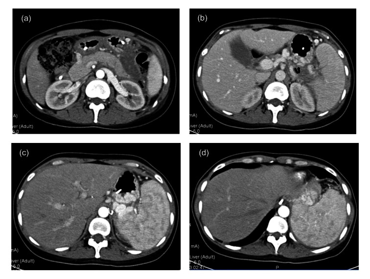
Fig. 1.
Enhanced computed tomography (CT) images of the pancreas of the patient
(a) An edematous area around pancreas with peripancreatic fat stranding and fluid (black arrow); (b, c) Severe, tortuous gastric (black curved arrow) and perisplenic varices (white curved arrow) were present; (d) The spleen was extremely enlarged without sign of splenic vein thrombosis (SVT)

