Figure 4.
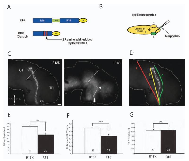
Inhibition of 14-3-3/14-3-3ς shortens retinal axon projection in vivo. (A) Schematics of R18 and R18K constructs tagged with YFP. (B) Schematics of eye electroporation. FITC-tagged morpholinos are electroporated into the eye at stage 26. (C) Dissected brains from stage 40 embryos that have been electroporated with either R18-YFP or R18K-YFP. The arrowhead indicates markedly shorter axons found in R18-YFP transfected brains (dashed line = anterior tectal border; scale bar = 50 μm; CH = optic chiasm; TEL = telencephalon; OT = optic tectum). (D) The length of the pathway was measured in two ways: (1) following the curvature of the pathway (a) and (2) measuring the shortest distance from the optic chiasm to the longest axon (LA; b) divided by the shortest distance from the chiasm to the posterior tectum (PT; c). (E) Measurement of the pathway length (equivalent to “a” in Fig. 4D). (F) Measurement of CH to LA length/CH to PT length (equivalent to “b/c” in Fig 4.4). (G) Measurement of CH to PT length to rule out significant differences in brain size. (Note: in E, F, and G, n = number of brains analyzed; three replicates; **p<0.01; ***p<0.001; unpaired t-test).

