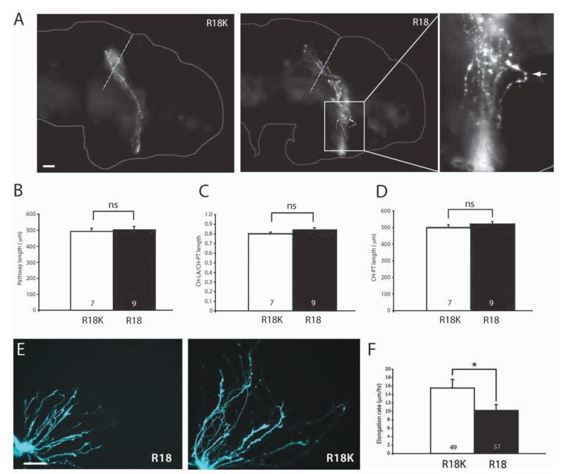
Figure 5.
Shortened retinal projection due to retarded axon elongation rate. (A) Brains of stage 43 embryos that have been eye-electroporated with R18-YFP or R18K-YFP (R1). Axons reach the tectum even in those that were electroporated with R18-YFP. The rightmost panel represents the magnified view of the R18 pathway with an axon taking an unusually tortuous path (arrowhead; dashed line = anterior tectal border; scale bar = 50 μm). (B) Measurement of the pathway length. (C) Distance from CH-LA/CH-PT. (D) Distance from CH-PT (For B, C, D, n = no. of brains analyzed; unpaired t-test). (E) Whole eye culture of eyes that have been electroporated with R18-YFP or R18K-YFP (scale bar = 50 μm). (F) The elongation rate of axons transfected with R18-YFP or R18K-YFP (n = no. of growth cones; *p<0.05; Mann–Whitney). [Color figure can be viewed in the online issue, which is available at wileyonlinelibrary.com.]

