Figure 6.
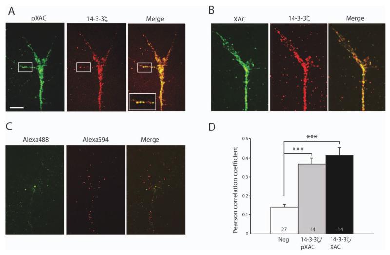
Colocalization of 14-3-3ς with pXAC and XAC. (A) Growth cone staining with Alexa 488-conjugated pXAC antibody and Alexa 594-conjugated 14-3-3ς antibody. The squared area shows colocalization in a filopodium. The inset shows more magnified view of the squared area (scale bar = 5 μm; image intensity adjusted). (B) Growth cone staining with Alexa 488-conjugated XAC antibody and Alexa 594-conjugated 14-3-3ς antibody. (C) Growth cone staining with Alexa 488 and Alexa 594 fluorophores without antibody conjugation. (D) Measurement of Pearson coefficient for determination of colocalization (n = no. of growth cones;***p<0.001; statistical analysis by Kruskal–Wallis test with Dunn post-test). (R1)

