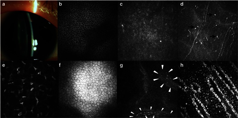
Figure 3.
Clinical findings in case 3. (a) Slitlamp biomicroscopic photo shows a clear cornea. (b–f) Confocal microscopic images of the central cornea. The morphology of wing and basal epithelial cells was fairly normal (b, c). The subbasal nerve plexus were visible, along with the infiltration of dendritic cells (black arrows) (d). The morphology of corneal stroma and endothelium was normal (e, f). (g, h) Confocal images of the limbus. The palisade-like stromal cords were identified at limbus of this patient (white triangles) (g). Within the lower cord in the image, a slender blood vessel was visible. Panel (h) was the image taken from an age-matched healthy subject, revealing that hyperreflective cells were present around stromal cords in normal Vogt palisades.

