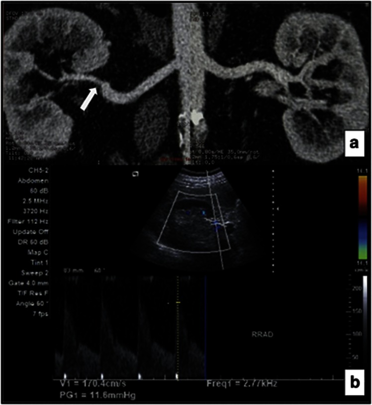
Figure 1.
A 64-year-old male patient with significantly stenotic right renal artery in PEX group. (a) CT angiography demonstrated a stenosis (arrow) of the distal right renal artery. The left renal artery was normal. (b) Direct sonographic examination of distal right renal artery shows PSV of 170 cm/s, indicating significant stenosis.

