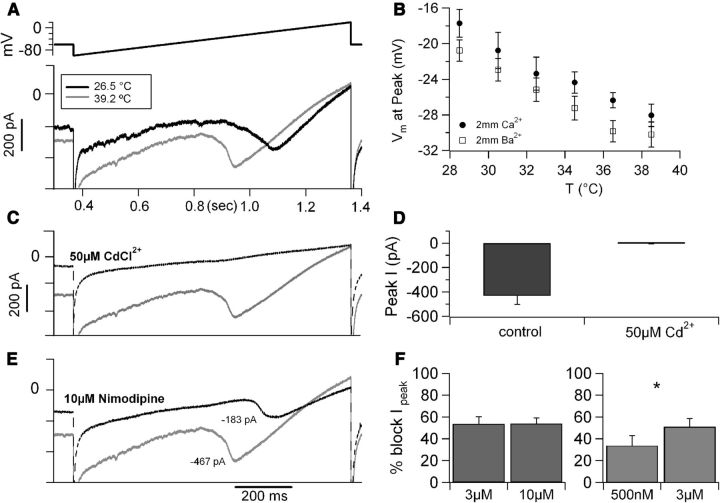
Figure 6.
Temperature modulates L-type calcium current in CA1 neurons. A, Whole-cell current recording of a CA1 pyramidal cell at 26.5°C (black trace) and at 39.2°C (gray trace) in response to a slow voltage ramp from −100 to +20 mV (0.12V/s, top trace). B, The voltage at which the current peaked plotted versus recording temperature. These data were obtained from recordings performed in extracellular solution containing 2 mm Ca2+ (n = 6) or 2 mm Ba2+ (n = 11); in both conditions, temperature increase induced a shift in the hyperpolarizing direction of ∼1mV/°C. C, The temperature-sensitive current was carried by calcium ions, because it was blocked by CdCl2 (50 μm, dotted trace). D, Average peak current measured at high temperature (38.6 ± 0.2°C, 6 cells) in 2 mm external Ba2+ in control conditions and in the presence of 50 μm CdCl2. E, Current recordings obtained at 39.2°C in control conditions (gray trace) and after focal application of 10 μm nimodipine (black dotted trace). The traces in A, C, and E were obtained from the same neuron with sequential drug applications. F, Left, Different concentrations of nimodipine (3 μm, n = 4 and 10 μm, n = 5, applied focally at 35.4–39.3°C) induced similar current block. Right, In a different set of recordings, 500 nm nimodipine blocked ∼65% of the nimodipine-sensitive current (defined as the current blocked by 3 μm nimodipine in the same cells, n = 3, recorded at 36.1 ± 0.5°C). *p < 0.05.

