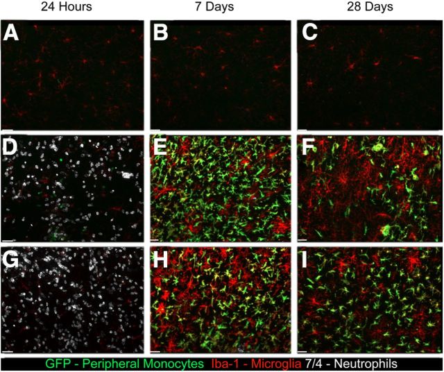
Figure 4.
URMC-099 does not alter the peripheral cellular immune response to cortical HIV-1 Tat injection. We performed bone morrow transplantation in lethally irradiated 8-week-old CD45.1 mice with CX3CR1/GFP+/− donor bone marrow and allowed the mice to recover for 4 weeks. We then injected either 3 μl of sterile PBS (A–C) or 3 μl of 3 μg/μl HIV-1 Tat 700 μm deep into somatosensory cortex. The mice receiving Tat were either not treated (D–F) or were treated with intraperitoneal injections of 10 mg/kg URMC-099 twice a day before and after Tat exposure (G–I). We killed the mice 24 h, 7 d, and 28 d after Tat injection and collected the tissue for immunohistochemical analysis. We stained for the neutrophil marker 7/4 (white), GFP (green), which was present only in monocytes derived from the peripheral bone marrow, and the monocyte/microglia marker Iba-1 (red). Images shown were acquired using a 20× air objective and are shown as an accumulative z-projection of a 16 μm z-stack with a 0.5 μm z-step. Scale bars, 32 μm.

