
Figure 6.
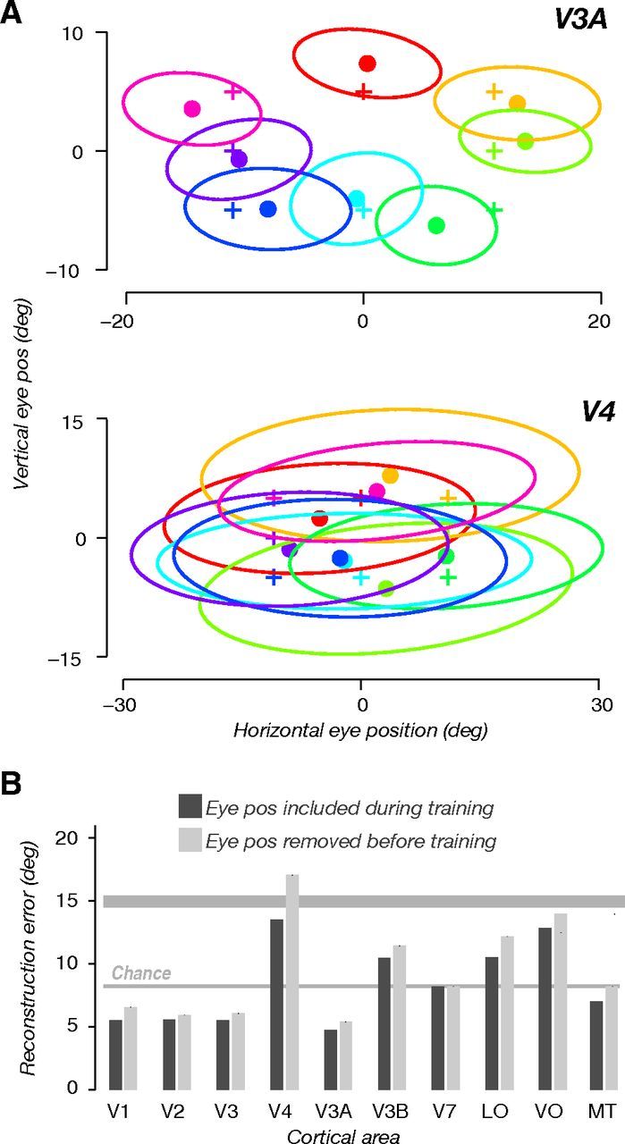
Planar gain field model enables accurate eye position reconstruction. A, Reconstructed eye position from response amplitudes in V3A (top) and V4 (bottom). Data pooled across subjects and sessions. +, actual eye position; ·, median reconstructed eye position; ellipses, 1 SD. B, Error in reconstructed eye position (median distance between reconstructed and actual eye position). Smaller values mean better reconstruction (opposite to Fig. 4). Dark bars, eye position reconstruction error when all eight eye positions were included in training to fit the planar gain field model. Light bars, eye position reconstruction error when the tested eye positions were excluded (one at a time) from training/fitting. Thick and thin horizontal gray lines indicate the error that would have been achieved by chance: 50th and 5th percentile, respectively, of the null distribution, according to the null hypothesis that eye position could not be reconstructed.