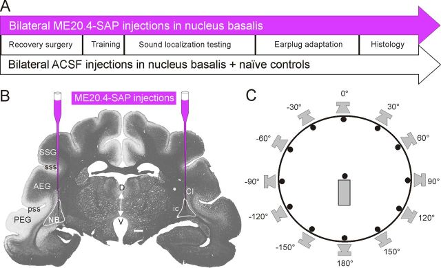
Figure 1.
Overview of experimental procedures. A, Time lines for the two main experimental groups used in this study. Injections of ME20.4-SAP or ACSF in the NB were performed before behavioral training. Subsequently, animals were tested for their ability to localize sounds in azimuth both under normal hearing conditions and then in the presence of a unilateral earplug, before perfusion and histology. B, Photograph of a coronal section of a ferret brain, stained for myelin using the Gallyas method, at the level of the auditory cortex and the NB, illustrating the position of ME20.4-SAP immunotoxin injections. C, Ferrets were trained by positive conditioning to approach the perceived location of an auditory stimulus presented from 1 of 12 speakers arranged around the periphery of a circular chamber. Animals initiated trials by standing on a central platform and licking the associated spout for at least 300 ms. Scale bar (in B), 1 mm. AEG, Anterior ectosylvian gyrus; Cl, claustrum; D, dorsal; ic, internal capsule; PEG, posterior ectosylvian gyrus; pss, pseudosylvian sulcus; SSG, suprasylvian gyrus; sss, suprasylvian sulcus; V, ventral.

