
Figure 8.
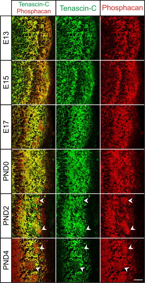
Colocalization of Tnc and Phosphacan in the developing OB. Coronal section of OB from E13–E17 and PND0–PND4 stained with Tnc (green) and phosphacan (red). Tnc and phosphacan partially colocalize, as evident by yellow overlap. However, distinct red and green staining is also evident. Phosphacan is found more extensively within glomerular structures than TNC (arrowheads). Both proteins are in a position to influence OSN axon growth. Scale bar, 50 μm.