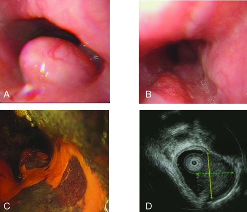
Figure 1.

Endoscopic views of the small cell carcinoma of esophagus. A) A submucosal elevated lesion (1.5x1.5 cm) covered with normal esophageal mucosa at 25 cm in esophagus; B) a patch-like erosion is observed at 24-28 cm (6:00-7:00 position) in esophagus before staining; C) a large irregular unstained area at 24-28 cm in esophagus after Lugol's iodine staining; D) a homogeneous, isoechoic mass (1.0×1.1 cm2) with a regular border originating from submucosal layer in esophagus, which is diagnosed as a granular cell tumor by EUS.
