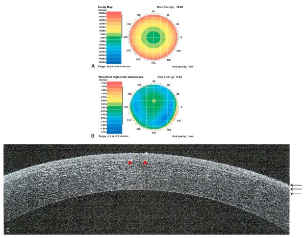
FIGURE 1.
Custom LASIK after 1 year. Preoperative acuity map of patient with manifest refraction of −10.71 +1.36 × 81 (A). Preoperative wavefront high-order aberrations of a patient with defocus of 14.8, astigmatism of 0.90, coma of 0.205, trefoil of 0.10, and spherical aberration of 0.27 (B). Spectral-domain optical coherence tomography showing a cross-section of the cornea. Red arrow-heads showing the LASIK flap (C). Three black arrows showing the epithelium, Bowman’s membrane, and stroma, respectively.

