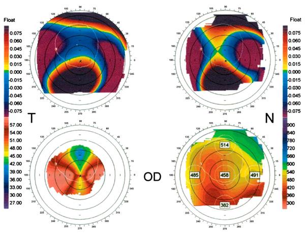
FIGURE 2.
Claw-shaped topography patterns in patients with PMD. Orbscan II topographic data of a representative patient with PMD. (Upper left) Anterior elevation float. (Upper right) Posterior elevation float. (Lower left) Mean keratometric axial power map showing superior flattening and horizontal steepening. (Lower right) Pachymetry map. Note the thin pachymetry in the inferior corneal periphery. Reprinted with permission from Lee et al.14

