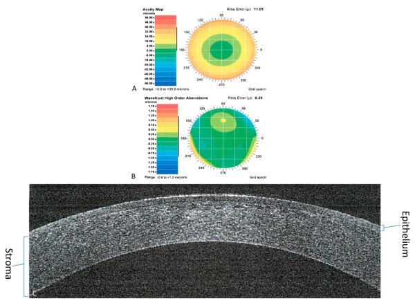
FIGURE 4.
One year after custom PRK. Preoperative acuity map of a patient with manifest refraction of 11.54 + 1.81 × 96 (A). Preoperative wavefront high-order aberrations of a patient with defocus of 10.96, astigmatism of 0.96, coma of 0.16, trefoil of 0.14, and spherical aberration of 0.046 (B). Spectral-domain optical coherence tomography showing a cross-section of the cornea showing the healed epithelium (C).

