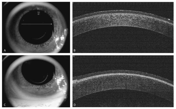
FIGURE 5.
Spectral-domain optical coherence tomography immediately after Epi-LASIK. Image taken from the center of the cornea (white arrow) after application of a therapeutic nonrefractive soft contact lens (A), with its corresponding cross-section of the cornea (B). Image taken from the inferior edge of the cornea (white arrow) after application of a therapeutic nonrefractive soft contact lens (C), with its corresponding cross-section of the cornea (D).

