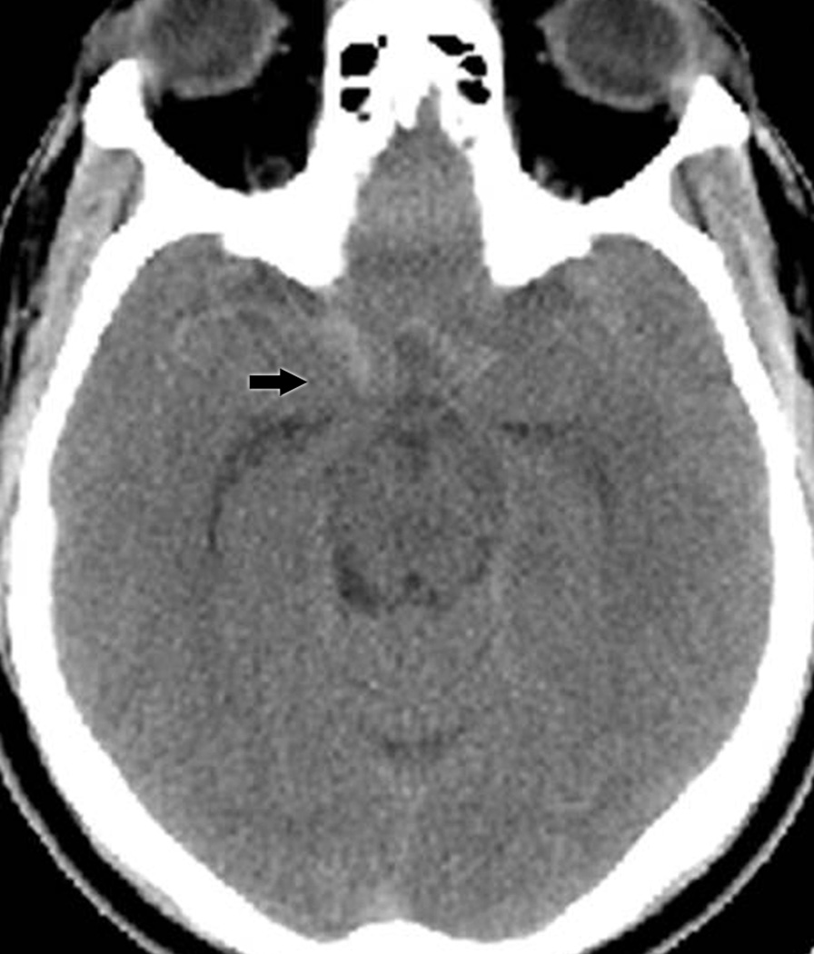
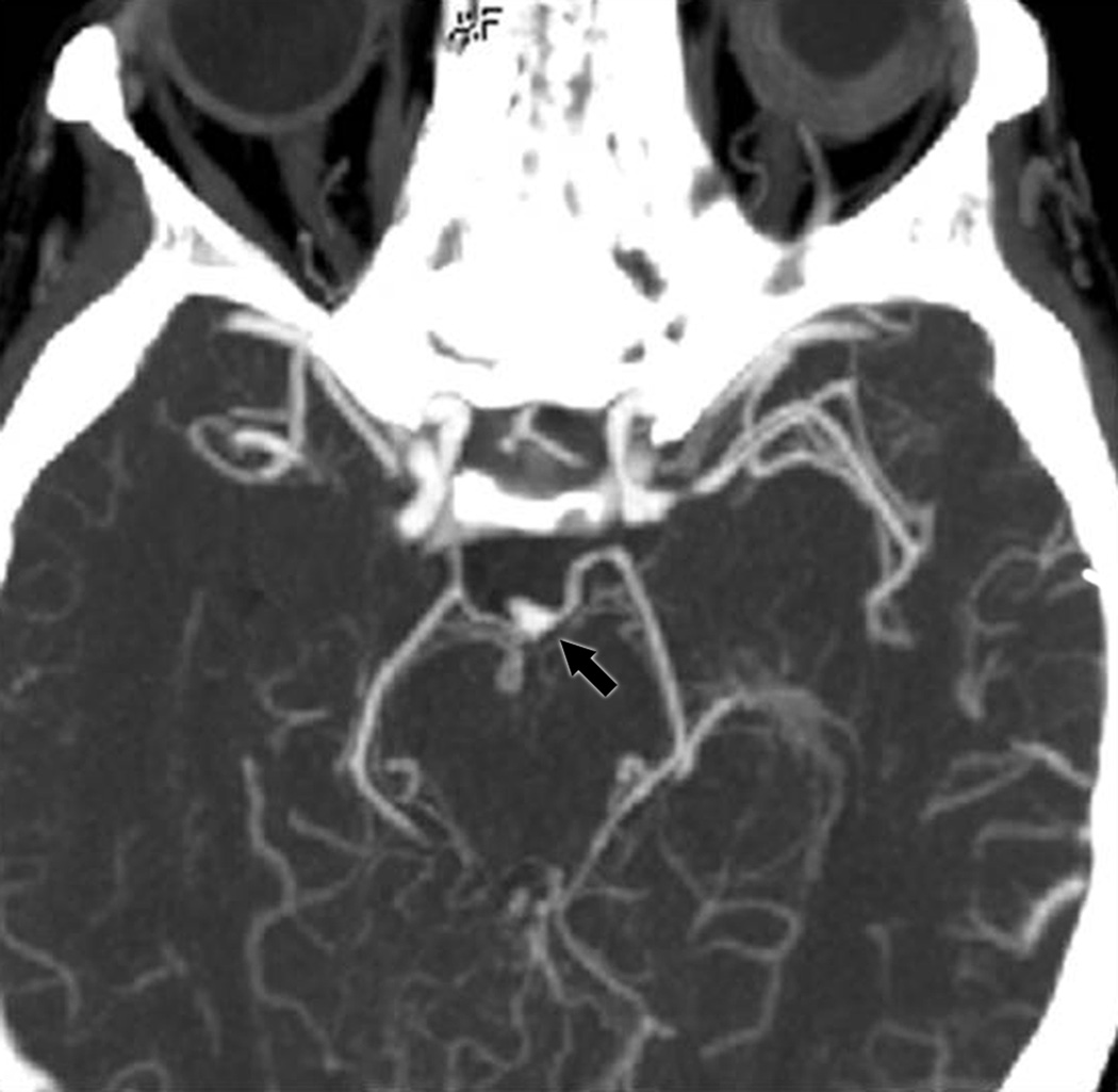
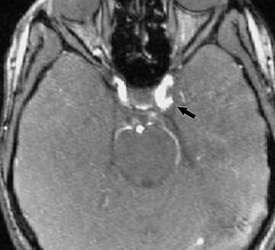
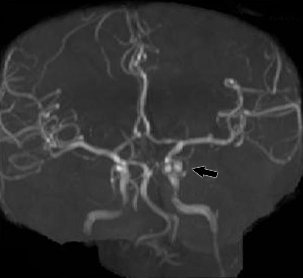
Figure 1.
A: Non-contrast head CT showing subarachnoid hemorrhage (arrow) in a patient with an isolated painful right third nerve palsy (Group I).
B: CTA demonstrating a left posterior communicating artery aneurysm (arrow) in a patient from Group II.
C: (top) MRA reformatted image from a patient in Group III showing a posterior communicating artery aneurysm (arrow); (bottom) MRA source image from the same patient showing the aneurysm (arrow).


