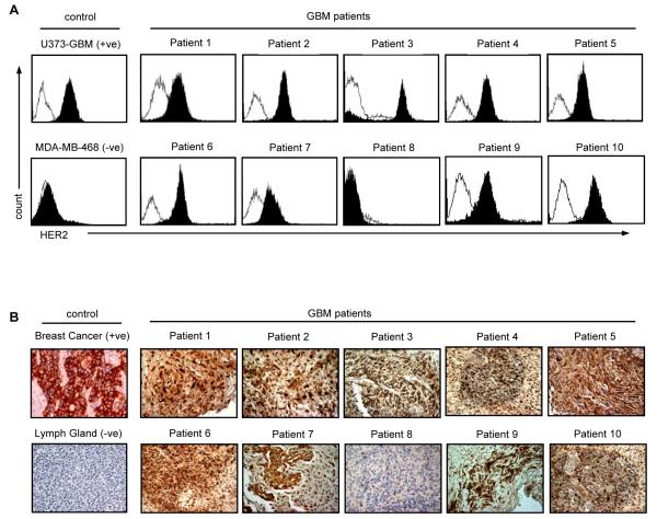
Figure 2. HER2 protein expression on primary GBM.
(A) FACS analysis: primary GBM cells from freshly excised tumors in short term culture were stained for HER2 expression (isotype control: open curves; HER2: solid curve. Nine out of ten tumor cell lines expressed HER2 on the cell surface. (B) Using the HER2-specific mouse monoclonal antibody NCL-L-CB11 (NovocastraTM Newcastle upon Tyne, UK), HER2 expression was confirmed on the corresponding paraffin-embedded sections. One tumor had no detectable HER2 protein expression using both methods (patient 8). Magnification 200x.

