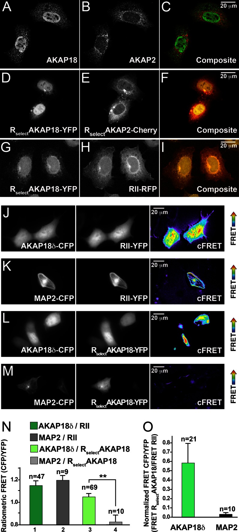
FIGURE 4.
RSelect subunits exhibit AKAP selectivity in cells. A–I, confocal images of full-length AKAPs and RSelect D/D domain fragments show their subcellular location. Immunofluorescent images show HEK293 cells cotransfected with FLAG-AKAP18δ (A; green in C) and AKAP2-V5 (B; red in C). Fluorescent images show RSelectAKAP18-YFP (D; yellow in F) and RSelectAKAP2-Cherry (E; red in F), which were coexpressed with AKAP18δ and AKAP2. Fluorescent images show RSelectAKAP18-YFP (G; yellow in I) and RII-RFP (H; red in I), which were coexpressed with AKAP18δ. J–M, CFP-YFP FRET imaging of the following RII-AKAP or RSelectAKAP18 pairs in HEK293 cells: AKAP18δ-CFP and RII-YFP (J), MAP2-CFP and RII-YFP (K), AKAP18δ-CFP and RSelectAKAP18-YFP (L), and MAP2-CFP and RSelectAKAP18-YFP (M). Images were acquired for donor CFP (left panels) and acceptor YFP (middle columns), and corrected FRET images are presented (right panels). N, ratiometric quantification of intermolecular FRET pairs as described for J–M. **, p < 0.01. O, quantification of FRET signals in AKAP18δ-CFP/RSelectAKAP18-YFP and MAP2-CFP/RSelectAKAP18-YFP pairs normalized to AKAP18δ-CFP/RII-YFP and MAP2-CFP/RII-YFP, respectively.

