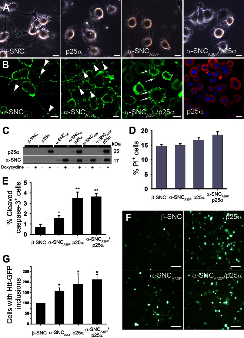
FIGURE 1.
Conditional expression of p25α and α-synuclein in NGF-differentiated PC12 nerve cells. PC12 cells were predifferentiated with 100 ng/ml NGF for 2 days, and then transgene expression of β-synuclein (β-SNC), p25α, α-synucleinwt (α-SNCwt), α-synucleinA30P (α-SNCA30P), or α-SNCA30P and p25α was induced by doxycycline treatment for additionally 2 days. A, bright field images showing p25α-mediated impairment of neurite outgrowth. Bars, 20 μm. B, indirect immunofluorescence of PC12 cells expressing α-SNCwt, α-SNCA30P, α-SNCA30P/p25α, or p25α alone with antibodies against α-SNC (BD Transduction Laboratories) (green) or p25α (red). Arrowheads indicate neurite blebbing, and arrows indicate α-SNC-positive inclusions. Bars, 20 μm. C, representative Western blots of transgene expression in doxycycline-treated or -nontreated PC12 cell lines analyzed with antibodies against p25α and α-SNC (BD Transduction Laboratories) as indicated. D, flow cytometry analysis of PI uptake as a measurement of cell death. The graph shows mean ± S.E. of PI-positive cells as a percentage of the whole population (n = 3). E, quantitation of caspase-3-positive cells detected by indirect immunofluorescence and counting. The bar graph shows mean ± S.E. values as percentage caspase-3-positive cells of the whole population (n = 3). F, differentiated PC12 cells lines were transduced with a lentivector expressing a pathogenic polyglutamine tract from exon 1 of huntingtin fused to GFP (Htt-115Q-GFP) and then induced with doxycycline. The images show microscopic fields containing ∼50 cells, whereof a proportion is highly fluorescent due to inclusion body formation. Bar, 100 μm. G, bar graph shows the number of PC12 cells per microscopic field containing Htt-115Q-GFP inclusion bodies after 4 days, normalized to PC12 cells co-expressing β-synuclein (β-SNC). The data represent mean ± S.E. from three independent experiments.

