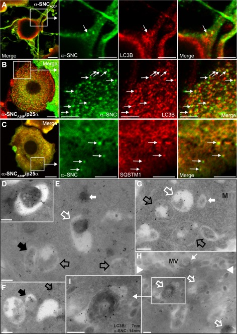
FIGURE 3.
Autophagy markers LC3B and p62/SQSTM1 co-localize with α-synuclein. Indirect immunofluorescence of PC12 cells expressing α-synucleinA30P alone (α-SNCA30P) (A) or together with p25α (B and C) to visualize α-SNC (mAb LB509) distribution in relation to LC3B (A and B) or p62/SQSTM1 (C). Co-localization between α-SNC and the respective markers are indicated with arrows. Bars, A–C, 10 μm in the left panels and 5 μm in close-ups. PC12-α-SNCA30P (D–F) or PC12-α-SNCA30P/p25α (G–I) cells expressing mCherry-eGFP-LC3B were fixed and processed for cryo-immunogold labeling with mouse monoclonal anti-α-SNC (mAb LB509) antibodies and rabbit polyclonal anti-GFP antibodies, followed by secondary 14- or 7-nm gold-conjugated anti-mouse or anti-rabbit antibodies, respectively. E, micrograph shows several autophagosomes containing only labeling for eGFP-LC3B (black open arrows) and a single autophagosome containing both α-SNC and eGFP-LC3B (black filled arrow), which is also true of an electron dense autolysosome (open white arrow). The closed white arrow points to a cytosolic inclusion staining for both α-SNC and eGFP-LC3B. D, autolysosome with both labels is also shown at higher magnification. F, nascent autophagosome with both labels (black open arrow) and a vacuole/autophagosome containing only aggregated label for α-SNC (black filled arrow). In PC12 cells co-expressing α-SNCA30P and p25α (G–I), the number of autophagosomes was increased and recently formed autophagosomes (open black arrows) and amphisomes (white open arrows), including lamellar bodies (white, open arrow in G) and electron dense late autophagosomal elements (white, open arrows in H) contained label for both α-SNC and eGFP-LC3B. White closed arrow in G points to a late endosome devoid of immunoreactivity for either α-SNC or eGFP-LC3B, and the small white arrow in H points to extracellular α-SNC immunoreactivity associated with microvilli (MV) on the cell surface (white arrowheads). I, electron dense late autophagosomal element from H shown at higher magnification. M, mitochondria. Bars, D–H, 500 nm.

