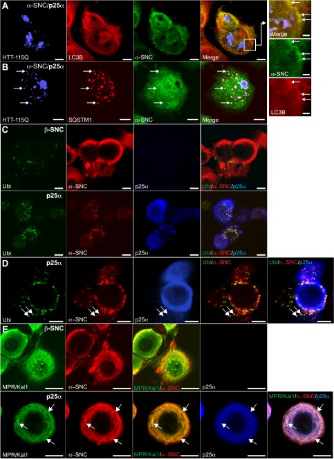
FIGURE 4.
TPPP/p25α-induced autophagy is selective and also affects endogenous rat α-synuclein. A and B, PC12 α-synucleinA30P/p25α (α-SNCA30P/p25α) cells were transduced with lentivector pHR-cPPT.CMV.W-Htt-115Q-GFP, and after 2 days α-SNC and p25α expression was induced with doxycycline for a further 2 days. Cells were then fixed and processed for indirect immunofluorescence to visualize the following: A, Htt-115Q-GFP (blue), LC3B (red), and α-SNC (green; BD Transduction Laboratories), or B, Htt-115Q-GFP (blue), p62/SQSTM1 (red), and α-SNC (green; BD Transduction Laboratories). The box in A is shown at higher magnification to the right. Note that α-SNC preferentially co-localizes with LC3B, whereas Htt-115Q-GFP co-localizes with p62/SQSTM1. Bars, 10 μm and in right panels 2 μm. C, NGF-differentiated PC12 cells expressing either β-synuclein (β-SNC) as control or p25α were analyzed by indirect immunofluorescence to localize polyubiquitin, endogenous α-SNC (Abcam mAb EP1646Y), and p25α as indicated. D, PC12 cells expressing p25α and stained as above at higher magnification. Arrows indicate co-localization of α-SNC with ubiquitin and p25α. E, control (β-SNC) or p25α-expressing PC12 cells were processed for indirect immunofluorescence with rat anti-p25α mAb, mouse anti-α-SNC mAb LB509, and rabbit polyclonal antibodies against KAI1 and MPR. Arrows indicate vesicular structures with co-localization of α-SNC and KAI1/MPR, which to a certain extent also co-localize with p25α. Bars, C–E, 10 μm.

