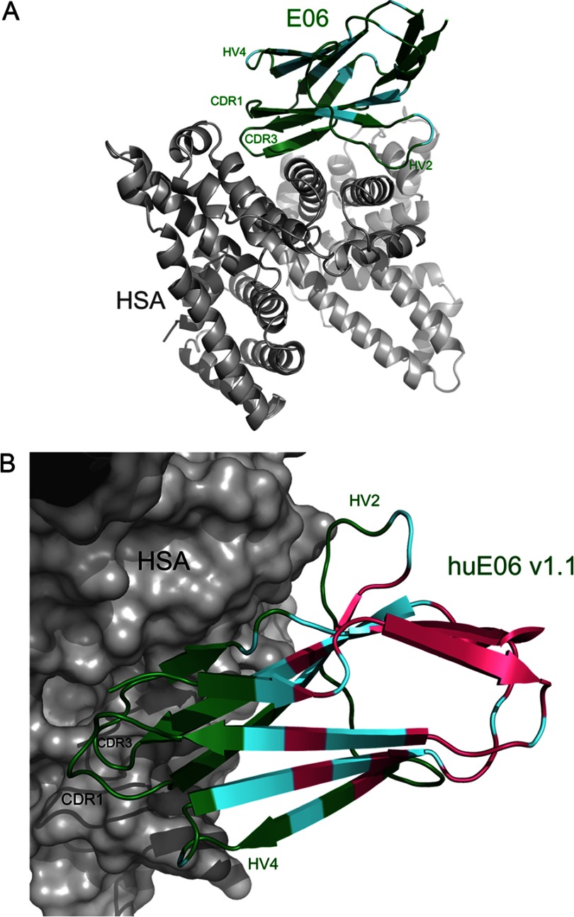
FIGURE 3.
Ribbon diagrams of E06 and huE06 v1.1 in complexes with HSA. E06 and huE06 v1.1 are shown in color, and HSA is in gray. V-NARs are colored according to the following: residues with the original shark sequence are shown in green, residues in which the original shark sequence are identical to human germ line DPK9 are shown in cyan, and residues that have been mutated from the original shark sequence to correspond to DPK9 are colored in pink. A, the structure of the E06-HSA complex. B, A closer view of the huE06 v1.1 structure bound to HSA with HSA shown as a surface representation.

