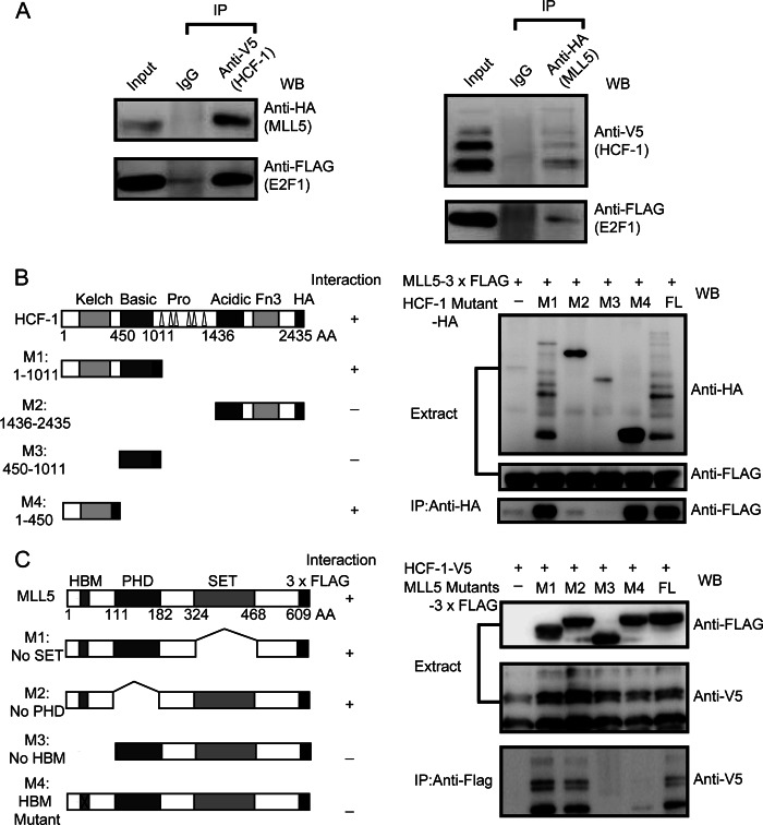
FIGURE 2.
Mapping of interacting domains of MLL5 and HCF-1 protein. A, shown is the association of MLL5, HCF-1, and E2F1 proteins. HEK293T cells were co-transfected with plasmids encoding HA-tagged MLL5, V5-tagged HCF-1, and FLAG-tagged E2F1. Left panel, V5-tagged HCF-1 protein was immunoprecipitated (IP) with anti-V5 antibody, and the presence of HA-tagged MLL5 and FLAG-tagged E2F1 protein were examined by Western blotting using anti-HA and anti-FLAG antibodies, respectively. Right panel, HA-tagged MLL5 protein was immunoprecipitated with anti-HA antibody, and the presence of V5-tagged HCF-1 and FLAG-tagged E2F1 proteins were examined by Western blotting using anti-V5 and anti-FLAG antibodies, respectively. B, mapping the MLL5 interacting domain in the HCF-1 protein. Left panel, a schematic representation of the domain structures of the full-length HCF-1 protein and truncated mutants. Right panel, HEK293T cells were co-transfected with plasmids encoding 3× FLAG-tagged MLL5 and HA-tagged full-length HCF-1 and truncated mutants; HA-tagged HCF-1 and truncated mutants were co-immunoprecipitated with anti-HA antibody, and the presence of 3× FLAG-tagged MLL5 protein was examined by Western blotting using anti-FLAG antibody. C, mapping the HCF-1 interacting domain in MLL5 protein. Left panel, a schematic representation of the domain structures of full-length MLL5 and truncated mutants. Right panel, HEK293T cells were co-transfected with plasmids encoding HA-tagged HCF-1 and 3× FLAG-tagged full-length MLL5 and truncated mutants, 3× FLAG-tagged MLL5 and truncated mutants were co-immunoprecipitated with anti-FLAG antibody, and the presence of HA-tagged HCF-1 protein were examined by Western blotting using anti-HA antibody.

