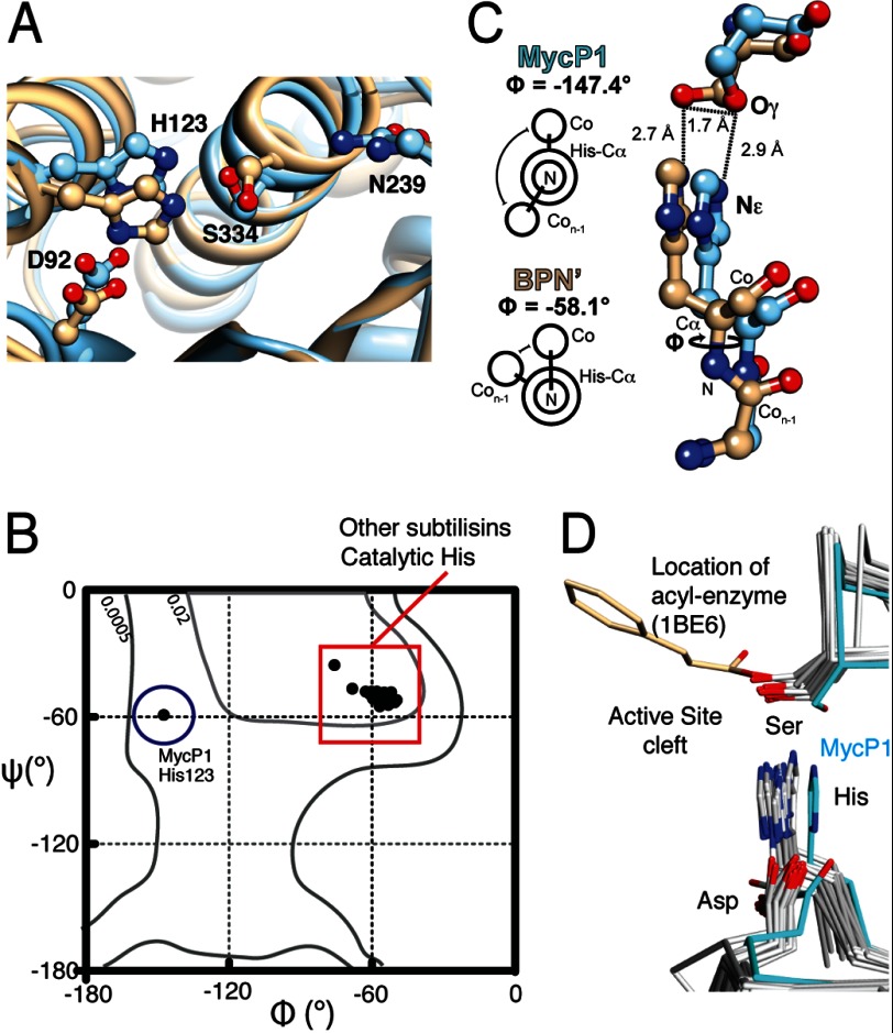
FIGURE 3.
MycP1 catalytic triad. A, structural overlay of MycP1 (blue) and subtilisin BPN′ (PDB ID code 2SNI, tan) with catalytic triads displayed. B, Ramachandran plot indicating ϕ/ψ torsion angles for MycP1 and 22 subtilisin-like enzymes. C, differing ϕ angles and His/Ser geometries for MycP1/subtilisin BPN′. D, structural alignment of subtilisin-like enzymes with catalytic triad displayed. The MycP1 triad is highlighted in cyan. An acyl-enzyme structure of subtilisin Carlsberg (PDB ID code 1BE6) indicates the serine location and orientation required for nucleophilic attack. Previously published PDB files used in structural alignment were: 1BE6, 3TI9, 3T41, 3LPC, 3F7M, 3EIF, 2Z30, 2Z2Z, 2QTW, 2IXT, 2B6N, 1Y9Z, 1TO2, 1THM, 1SH7, 1R6V, 1R0R, 1CGI, 1DBI, 1BH6.

