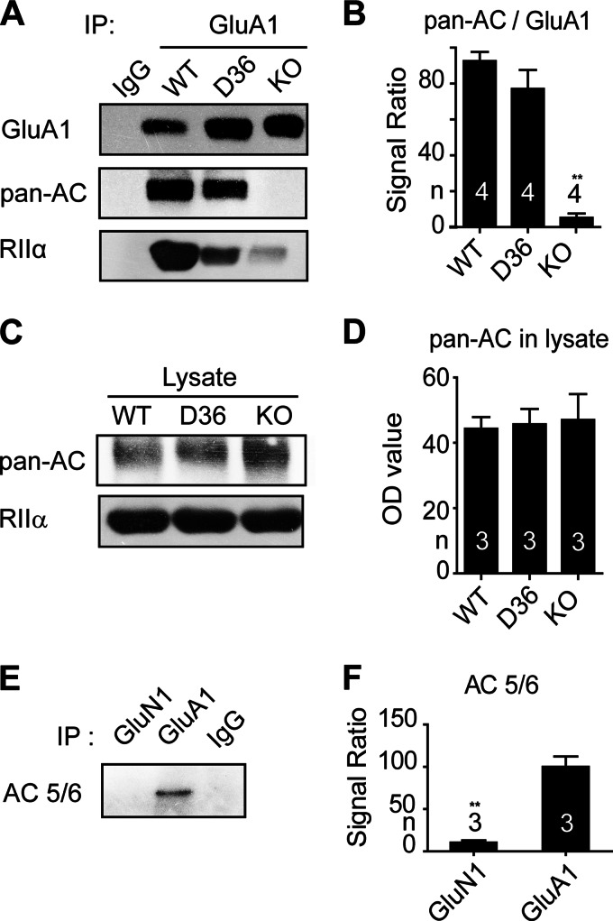
FIGURE 2.
AKAP5 links ACs to GluA1. Forebrains from WT, AKAP5 D36, and AKAP5 KO mice were extracted with Triton X-100 and cleared of non-solubilized material by ultracentrifugation. A, C, and E, lysate samples underwent IP with 1 μg of antibody against GluA1 or GluN1 or 1 μg of control rabbit IgG (A and E) or were directly applied (C) to immunoblotting with a panspecific antibody against all ACs and with antibodies against GluA1, RIIα, or AC5/6 as indicated. B, D, and F, immunosignals were quantified by densitometry. Depicted are film optical density (OD) ratios for pan-AC versus GluA1 signals (B), OD values for pan-AC (D), and OD ratios for AC5/6 signals in GluN1 versus in GluA1 IPs (F). Co-IP of ACs with GluA1 is nearly completely absent in GluA1 KO mice but not affected in D36 mice (**, p < 0.01, one-way ANOVA). Co-IP of AC5/6 with GluN1 is nearly undetectable compared with GluA1 IPs (**, p < 0.01, t test). Error bars, S.E.

