FIGURE 8.
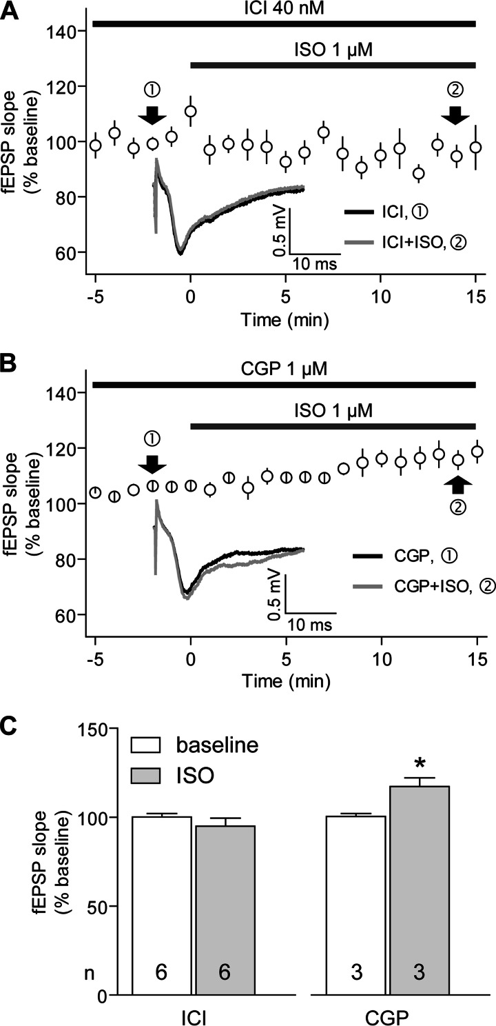
ISO-induced increases in basal synaptic transmission depend on β2-AR but not β1-AR. A and B, time courses of fEPSPs before and after perfusion with ISO (1 μm; bottom gray bar) in the presence of 40 nm ICI118551 (A) or 1 μm CGP20712 (B). Shown are averages of initial slopes of fEPSP starting after the base line had stabilized. Insets at bottom, examples of fEPSPs before (black lines) and after the start of ISO application (gray lines). Graphed are averages of 10 consecutive fEPSPs recorded at the indicated times (arrows). C, summary data. The base line is the average of the fEPSP initial slopes from each individual experiment during the 5 min immediately preceding the start of the ISO application and equals 100% for each experiment. The ISO bars show the increase in fEPSP responses, which were obtained by averaging the initial slope values measured 10–15 min after the onset of ISO perfusion. ISO did not induce any increase in fEPSPs in any of the six slices tested in the presence of ICI118551 (p = 0.3179; t test) but increased fEPSPs in the three slices tested in the presence of CGP20712 (*, p = 0.0297; t test). Error bars, S.E.

