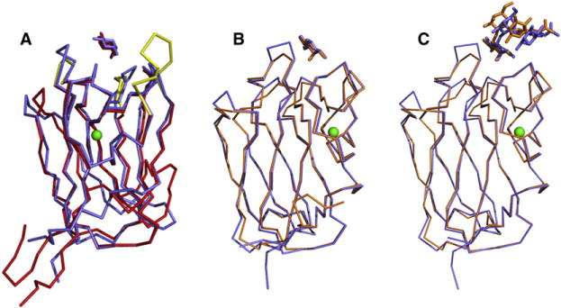
Figure 4. Structural Similarities of LLYlecto Other Proteins.

(A) Superposition of the LLYlec domain (in blue) on to the structure of the fucolection of AAA (in red). Both structures are complexed with fucose. The Ca2+ atom, which is located in the same positionin both structures, is shown in green. The CDR loops that are different in AAA, compared to the LLYLec domain, are shown in yellow.
(B) Superposition of the fucose complex structures of LLYlec domain (in blue) and the fucolectin module of SpX-1 (in orange).
(C) Superposition of the Ley antigen complex structures of LLYlec domain (in blue) and the fucolectin module of SpX-1 (orange).
