
Figure 2.
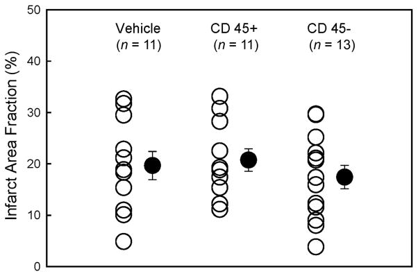
Myocardial infarct size. Myocardial infarct area fraction ([infarct area/left ventricular area] × 100) assessed from Masson’s trichrome-stained hearts in groups I–III, which were treated with vehicle, CD45+ hematopoietic stem cells, and very small embryonic-like stem cells, respectively. ○, individual mice; ●, mean ± SEM.