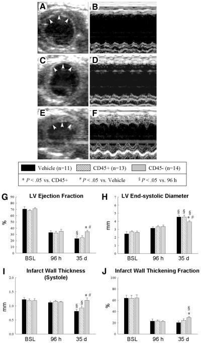
Figure 3.
Echocardiographic assessment of LV function. Representative two-dimensional (A, C, E) and M-mode (B, D, F) images from vehicle-treated (A, B), CD45+ cell-treated (C, D), and very small embryonic-like stem cell (VSEL)-treated (E, F) mice 35 d after coronary occlusion/reperfusion. The infarct wall is delineated by arrowheads (A, C, E). Compared with the vehicle-treated and CD45+ cell-treated hearts, the VSEL-treated heart exhibited a smaller LV cavity, a thicker infarct wall, and improved motion of the infarct wall. Panels (G–J) demonstrate that transplantation of VSEL improved echocardiographic measurements of LV systolic function 35 d after myocardial infarction. Data are mean ± SEM. n = 11–14 mice per group. *, p < .05 versus group II at 35 d; #, p < .05 versus group I at 35 d; §, p < .05 versus values at 96 h in respective groups. Abbreviations: BSL, baseline; d, days; h, hours; LV, left ventricular.

