Figure 4.
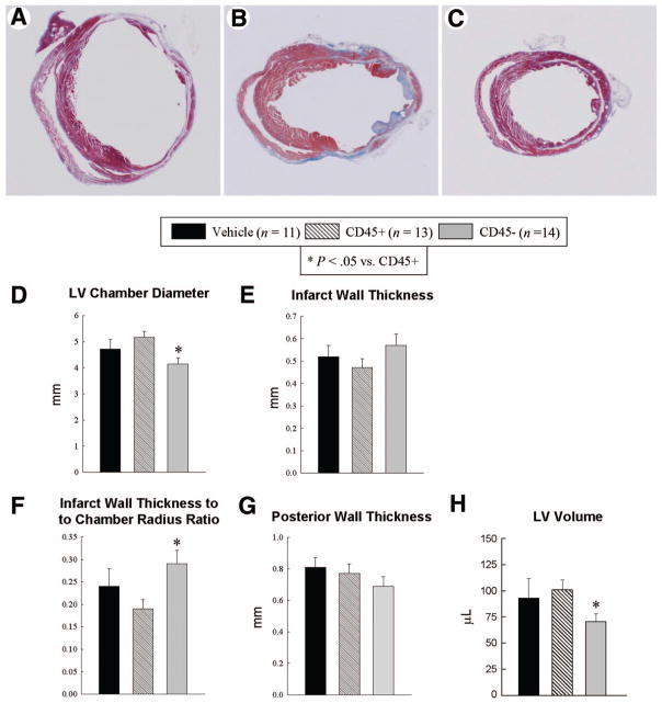
Morphometric assessment of LV remodeling. Representative Masson’s trichrome-stained myocardial sections from vehicle-treated (A), CD45+ hematopoietic stem cell-treated (B), and very small embryonic-like stem cell (VSEL)-treated (C) hearts. Scar tissue and viable myocardium are identified in blue and red, respectively. Note that the LV cavity is smaller and the infarct wall thicker in the VSEL-treated heart. Panels (D–H) illustrate morphometric measurements of LV structural parameters. Data are mean ± SEM. n = 11–14 mice per group. *, p < .05 versus group II. Abbreviation: LV, left ventricular.

