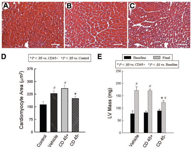
Figure 5.
Assessment of cardiomyocyte and LV hypertrophy. Panels (A–C) show representative images of cardiomyocytes in the viable myocardium from Masson’s trichrome-stained vehicle-treated (A), CD45+ hematopoietic stem cell-treated (B), and very small embryonic-like stem cell (VSEL)-treated hearts (C). Scale bars = 50 μm. In contrast to CD45+ hematopoietic stem cell-treated hearts, VSEL-treated hearts did not exhibit increased myocyte cross-sectional area compared with noninfarcted control hearts (D). Echocardiographically estimated LV mass was significantly less in VSEL-treated hearts (E). Data are mean ± SEM. n = 11–14 mice per group. (D): *, p < .05 versus group II; #, p < .05 versus control; (E): *, p < .05 versus group II and III (final); #, p < .05 versus respective baseline values. Abbreviation: LV, left ventricular.

