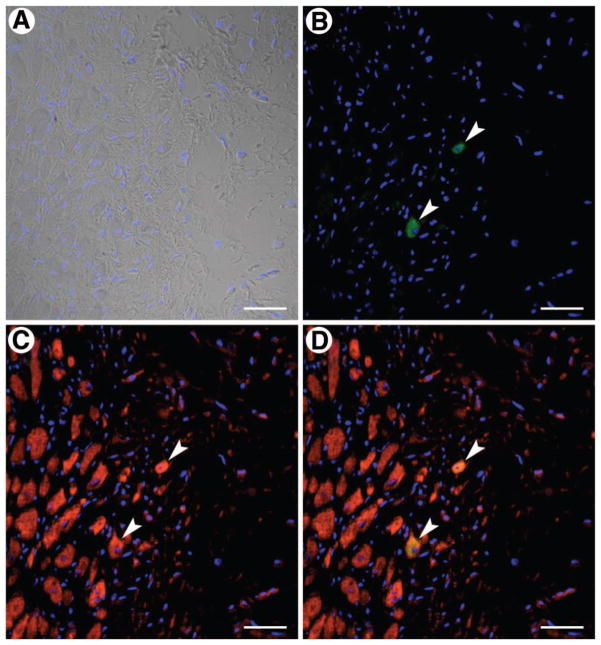
Figure 6.
Very small embryonic-like stem cell (VSEL) transplantation and cardiomyocyte regeneration. VSELs and myocytes are identified by enhanced green fluorescent protein (EGFP) ([B, D], green) and α-sarcomeric actin ([C, D], red), respectively; (D) shows the merged image. Two myocytes are shown that are positive for both EGFP (arrowheads; [B], green) and α-sarcomeric actin (arrowheads; [C], red). Nuclei were stained with 4,6-diamidino-2-phenylindole ([A, D], blue). Scale bars = 40 μm.

