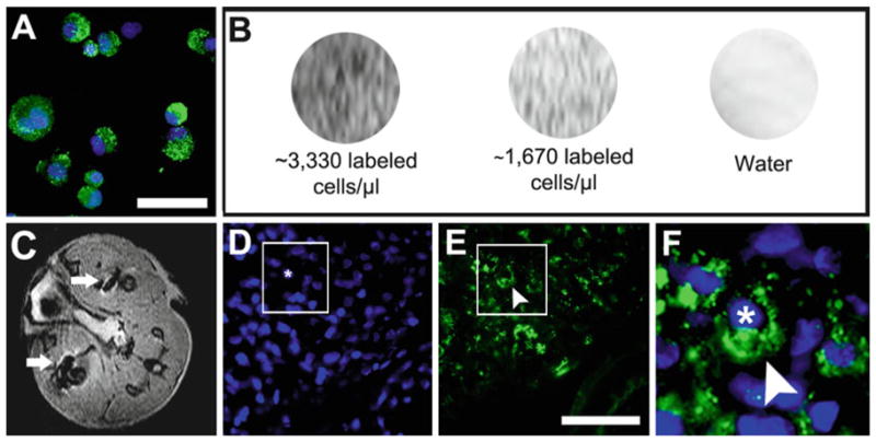
Fig. 4.

Detecting SPION labeled cells using MRI in vivo and in vitro. (a ) Appearance of FeProt labeled cells after trypsinization and immunolabeling for dextran. (b ) In vitro MRI of cells labeled for 4 h at different concentrations of cells/μL gelatin. (c) Representative image of in vivo MRI (transverse plane) showing hypointense (black) spots corresponding to ferumoxide-labeled cells injected in the leg muscles (white arrows). (d–f) Dextran immunocytochemistry confirming the presence of ferumoxide-labeled cells in sections of leg muscle; (d) nuclear counterstaining with DAPI; (e) dextran; (f) merged images in higher-magnification of the area indicated by the box in (d ) and (e ) showing a transplanted cell (nucleus marked with an asterisk) labeled with ferumoxide (arrowhead). Scale bar = 50 μm.
