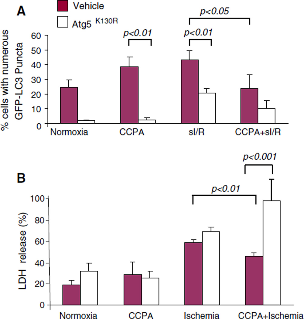
Fig. 9.
Role of autophagy in delayed preconditioning. HL-1 cells were co-transfected with GFP–LC3 and dominant negative Atg5K130R. Cells were treated with CCPA for 10 min, followed by washout. 20 h later, cells were subjected to sI/R (2 h sI, 3 h R). The extent of autophagy was assessed by the intracellular distribution of GFP-LC3 by fluorescence microscopy (a) and cell death was measured by LDH release into the medium at the end of ischemia (b)

